

Practical nursing not only for flunkers - Brion
Amid problems besetting the country’s nursing profession, Labor and Employment Secretary Arturo D. Brion on Wednesday said Licensed Practical Nursing (LPN) is an alternative course to nursing students who prefer to take the shorter route in their career and those who cannot make it in nursing board exams.
This would give them an opportunity to seek employment abroad, especially in countries like the United States and Canada that recognize and license practical nurses.
LPN nurses perform simple medical tasks, mostly dealing with patient medication and care, under the direction of a “full nurse" or a physician.
“We will have to find out what we can do for those who will not make it in the licensure examination and those who want to make the shorter LPN route in their career choice," Brion said.
He said the idea came up when he met recently with Senator Edgardo Angara to discuss the employment situation in the country, including the plight of the country's nurses.
Brion wanted the various stakeholders to debate on the suggestion, hoping that the ideas that would arise from the discussions could be consolidated into a legislative proposal.
"This is a policy issue that we will look into together with the stakeholders, particularly the Professional Regulation Commission and the Commission on Higher Education, and even the Technical Education and Skills Development Authority," Brion said.
The labor official said that of the 78,000 who took the examination last June, probably only 40,000 may pass based on passing rates in previous exams.
Referring to those who do not want to go through the full nursing course but still want to serve in the medical field and those who flunked in exams, Brion said, “We should provide them an option they can handle."
We do not license practical nurses in the Philippines, but they are recognized and licensed in the United States and Canada, Brion said.
The labor official noted media’s help in airing the idea, saying that public awareness could spark a discussion and debate on the issue.
"Our nurses and their organizations, together with the academe should be heard in this debate," he added.
The Board of Nursing expects to come out with the June 2007 nursing licensure exam results possibly by mid-August 2007.
FIRE HAZARD AND PREPAREDNESS PRACTICES
- Be aware of hazards and report immediately.
- Locate and remember
- Escape routes
- Fire drill procedures.
- Use of available equipment
- Fire escapes
- Fire doors
- Fire alarms
- Fire sprinkler controls
- Fire extinguishers
- Shut off valves for O2 and/or medical air
- Keep fire exits clear.
- Fire Safety
- Prevention is everyone's responsibility.
- Three elements needed for a fire to start
- Fuel--substance that will burn
- Heat--flame or spark
- Oxygen--room air contains 21% O2.
- See Table 3.3.
- In the event of a fire
- Move clients to safety if in immediate vicinity of fire
- Sound the alarm
- Close all windows and doors
- If selected clients need continuous O2 or medical air, attach to emergency provisions once they are removed from vicinity of fire
- Shut off piped-in O2 and/or medical air
- Follow institutional policy concerning announcing the fire and location and notifying fire company
- Avoid use of elevators
- Follow institutional evacuation plan as needed.
TABLE 3.3 Fire Hazards and Prevention
| Fire Hazards | Fire Prevention |
| Faulty electrical equipment and wiring | Report frayed or exposed electrical wires |
| Overloaded circuits | Avoid overloaded circuits |
| Plugs that are not properly grounded | Use only 3-pronged grounded plugs |
| Clutter | Avoid clutter |
| Unsafe practices when O2 in use | No open flames or smoking in the area |
| Smoking | Remove cigarettes and matches from room |
| Spontaneous combustion | Dispose of chemicals, rags, and combustible substances in proper containers |
RESPIRATORY SYSTEM
Health History
- Presenting problem
- Nose/nasal sinuses: symptoms may include colds, discharge, epistaxis, sinus problems (swelling, pain)
- Throat: symptoms may include sore throat, hoarseness, difficulty swallowing, strep throat
- Lungs: symptoms may include
- Cough: note duration; frequency; type (dry, hacking, bubbly, barky, hoarse, congested); sputum (productive vs nonproductive); circumstances related to cough (time of day, positions, talking, anxiety); treatment.
- Dyspnea: note onset, severity, duration, efforts to treat, whether associated with radiation, if accompanied by cough or diaphoresis, time of day when it most likely occurs, interference with ALD, whether precipitated by any specific activities, whether accompanied by cyanosis.
- Wheezing
- Chest pain
- Hemoptysis
- Life-style: smoking (note type of tobacco, duration, number per day, number of years of smoking, inhalation, related cough, desire to quit); occupation (work conditions that could irritate respiratory system [asbestos, chemical irritants, dry-cleaning fumes] and monitoring or protection of exposure conditions), geographical location (environmental conditions that could irritate respiratory system [chemical plants/industrial pollutants]); type and frequency of exercise/recreation.
- Nutrition/diet: fluid intake per 24-hour period; intake of vitamins
- Past medical history: immunizations (yearly immunizations for colds/flu; frequency and results of tuberculin skin testing); allergies (foods, drugs, contact or inhalant allergens, precipitating factors, specific treatment, desensitization)
Physical Examination
- Inspect for configuration of the chest (kyphosis, scoliosis, barrel chest) and cyanosis.
- Determine rate and pattern of breathing (normal rate 12-18/minute); note tachypnea, hyperventilation, or labored breathing pattern.
- Palpate skin, subcutaneous structures, and muscles for texture, temperature, and degree of development.
- Palpate for tracheal position, respiratory excursion (symmetric or asymmetric movement of the chest), and for fremitus.
- Fremitus is normally increased in intensity at second intercostal spaces at sternal border and interscapular spaces only.
- Increased intensity elsewhere may indicate pneumonia, pulmonary fibrosis, or tumor.
- Decreased intensity may indicate pneumothorax, pleural effusion, COPD.
- Percuss lung fields (should find resonance over normal lung tissue, note hyperresonance or dullness) and for diaphragmatic excursion (normal distance between levels of dullness on full expiration and full inspiration is 6-12 cm).
- Auscultate for normal (vesicular, bronchial, bronchovesicular) and adventitious (rales or crackles, rhonchi, pleural friction rub) breath sounds (see Figure 4.11).
FIGURE 4.11 Locations for hearing normal breath sounds 
Laboratory/Diagnostic Tests
- Arterial blood gases (ABGs)
- Measure base excess/deficit, blood pH, CO2, total CO2, O2 content, O2 saturation (SaO2), pCO2 (partial pressure of carbon dioxide), pO2 (partial pressure of oxygen)
- Nursing care
- If drawn by arterial stick, place a 4X4 bandage over puncture site after withdrawal of needle and maintain pressure with two fingers for at least 2 minutes.
- Gently rotate sample in test tube to mix heparin with the blood.
- Place sample in ice-water container until it can be analyzed.
- Pulmonary function studies
- Evaluation of lung volume and capacities by spirometry: tidal volume (TV), vital capacity (VC), inspiratory and expiratory reserve volume (IRV and ERV), residual volume (RV), inspiratory capacity (IC), functional residual capacity (FRC)
- Involves use of a spirometer to diagram movement of air as client performs various respiratory maneuvers; shows restriction or obstruction to air flow, or both.
- Nursing care
- Carefully explaining procedure will help allay anxiety and ensure cooperation.
- Perform tests before meals.
- Withhold medication that may alter respiratory function unless otherwise ordered.
- After procedure assess pulse and provide for rest period.
- Hematologic studies (ESR, Hgb and hct, WBC)
- Sputum culture and sensitivity
- Culture: isolation and identification of specific microorganism from a specimen
- Sensitivity: determination of antibiotic agent effective against organism (sensitive or resistant)
- Nursing care
- Explain necessity of effective coughing.
- If client unable to cough, heated aerosol will assist with obtaining a specimen.
- Collect specimen in a sterile container that can be capped afterwards.
- Volume need not exceed 1-3 ml.
- Deliver specimen to lab rapidly.
- Tuberculin skin test
- Intradermal test done to detect tuberculosis infection; does not differentiate active from dormant infections
- Purified protein derivative (PPD) tuberculin administered to determine any previous sensitization to tubercle bacillus
- Several methods of administration
- Mantoux test: 0.1 ml solution containing 0.5 tuberculin units of PPD-tuberculin is injected into the forearm.
- Tine test: a stainless steel disc with 4 tines impregnated with PPD-tuberculin is pressed into the skin.
- Results: read within 48-72 hours; inspect skin and circle zone of induration with a pencil; measure diameter in mm
- Negative: zone diameter less than 5 mm
- Doubtful or probable: zone diameter 5-10 mm
- Positive: zone diameter 10 mm or more
- Thoracentesis
- Insertion of a needle through the chest wall into the pleural space to obtain a specimen for diagnostic evaluation, removal of pleural fluid accumulation, or to instill medication into the pleural space
- Nursing care: pretest
- Confirm that a signed permit has been obtained.
- Explain procedure; instruct client not to cough or talk during procedure.
- Position client at side of bed, with upper torso supported on overbed table, feet and legs well supported.
- Assess vital signs.
- Nursing care: posttest
- Observe for signs and symptoms of pneumothorax, shock, leakage at puncture site.
- Auscultate chest to ascertain breath sounds.
- Bronchoscopy
- Insertion of a fiberscope into the bronchi for diagnosis, biopsy, specimen collection, examination of structures/tissues, removal of foreign bodies
- Nursing care: pretest
- Confirm that a signed permit has been obtained.
- Explain procedure, remove dentures, and provide good oral hygiene.
- Keep client NPO 6-12 hours pretest.
- Nursing care posttest
- Position client on side or in semi-Fowler's.
- Keep NPO until return of gag reflex.
- Assess for and report frank bleeding.
- Apply ice bags to throat for comfort; discourage talking, coughing, smoking for a few hours to decrease irritation.
THE ENDOCRINE SYSTEM
Health History
- Presenting problem: symptoms may include
- Change in appearance: hair, nails, skin (change in texture or pigmentation); change in size, shape, or symmetry of head, neck, face, eyes, or tongue
- Change in energy level
- Temperature intolerance
- Development of abnormal secondary sexual characteristics; change in sexual function
- Change in emotional state, thought pattern, or intellectual functioning
- Signs of increased activity of sympathetic nervous system (e.g., nervousness, palpitations, tremors, sweating)
- Change in bowel habits, appetite, or weight; excessive hunger or thirst
- Change in urinary pattern
- Life-style: any increased stress
- Past medical history: growth and development (any delayed or excessive growth); diabetes, thyroid disease, hypertension, obesity, infertility
- Family history: endocrine diseases, growth problems, obesity, mental illness
Physical Examination
- Check height, weight, body stature, and body proportions.
- Observe distribution of muscle mass, fat distribution, any muscle wasting.
- Inspect for hair growth and distribution.
- Check condition and pigmentation of skin; presence of striae.
- Inspect eyes for any bulging.
- Observe for enlargement in neck area and quality of voice.
- Observe development of secondary sex characteristics.
- Palpate thyroid gland (normally cannot be palpated): note size, shape, symmetry, any tenderness, presence of any lumps or nodules.
Laboratory/Diagnostic Tests
A variety of tests may be performed to measure the amounts of hormones present in the serum or urine in assessing pituitary, adrenal, and parathyroid functions; these tests will be referred to when appropriate under specific disorders of the endocrine system.
Thyroid Function
- Serum studies: nonfasting blood studies (no special preparation necessary)
- Serum T4 level: measures total serum level of thyroxine
- Serum T3 level: measures serum triiodothyronine level
- TSH: measurement differentiates primary from secondary hypothyroidism
- Radioactive iodine uptake (RAIU)
- Administration of 123I or 131I orally; measurement by a counter of the amount of radioactive iodine taken up by the gland after 24 hours
- Performed to determine thyroid function; increased uptake indicates hyperactivity; minimal uptake may indicate hypothyroidism
- Nursing care
- Take thorough history; thyroid medication must be discontinued 7-10 days prior to test; medications containing iodine, cough preparations, excess intake of iodine-rich foods, and tests using iodine (e.g., IVP) can invalidate this test.
- Assure client that no radiation precautions are necessary.
- Thyroid scan
- Administration of radioactive isotope (orally or IV) and visualization by a scanner of the distribution of radioactivity in the gland
- Performed to determine location, size, shape, and anatomic function of thyroid gland; identifies areas of increased or decreased uptake; valuable in evaluating thyroid nodules
- Nursing care: same as RAIU
Pancreatic Function
- Fasting blood sugar: measures serum glucose levels; client fasts from midnight before the test
- Two-hour postprandial blood sugar: measurement of blood glucose 2 hours after a meal is ingested
- Fast from midnight before test
- Client eats a meal consisting of at least 75 g carbohydrate or ingests 100 g glucose
- Blood drawn 2 hours after the meal
- Oral glucose tolerance test: most specific and sensitive test for diabetes mellitus
- Fast from midnight before test
- Fasting blood glucose and urine glucose specimens obtained
- Client ingests 100 g glucose; blood sugars are drawn at 30 and 60 minutes and then hourly for 3-5 hours; urine specimens may also be collected
- Diet for 3 days prior to test should include 200 g carbohydrate and at least 1500 kcal/day
- During test, assess the client for reactions such as dizziness, sweating, and weakness
- Glycosylated hemoglobin (hemoglobin A1c) reflects the average blood sugar level for the previous 100-120 days. Glucose attaches to a minor hemoglobin (A1c). This attachment is irreversible.
- Fasting is not necessary.
- Excellent method to evaluate long term control of blood sugar.
MUSCULOSKELETAL SYSTEM
Health History
- Presenting problem
- Muscles: symptoms may include pain, cramping, weakness
- Bones and joints: symptoms may include stiffness, swelling, pain, redness, heat, limitation of movement
- Life-style: usual patterns of activity and exercise (limitations in ADL, use of assistive devices such as canes or walkers), nutrition (obesity) and diet, occupation (sedentary, heavy lifting, or pushing)
- Use of medications: drugs taken for musculoskeletal problems
- Past medical history: congenital defects, trauma, inflammations, fractures, back pain
- Family history: arthritis, gout
Physical Examination
- Inspect for overall body build, posture, and gait.
- Inspect and palpate joints for swelling, deformity, masses, movement, tenderness, crepitations.
- Inspect and palpate muscles for size, symmetry, tone, strength.
Laboratory/Diagnostic Tests
- Hematologic studies
- Muscle enzymes: CPK, aldolase, SGOT (AST)
- Erythrocyte sedimentation rate (ESR)
- Rheumatoid factor
- Complement fixation
- Lupus erythematosus cells (LE prep)
- Antinuclear antibodies (ANA)
- Anti-DNA
- C-reactive protein
- Uric acid
- X-rays: detect injury to or tumors of bone or soft tissue
- Bone scan
- Measures radioactivity in bones 2 hours after IV injection of a radioisotope; detects bone tumors, osteomyelitis.
- Nursing care
- Have client void immediately before the procedure.
- Explain that client must remain still during the scan itself.
- Arthroscopy
- Insertion of fiberoptic endoscope (arthroscope) into a joint to visualize it, perform biopsies, or remove loose bodies from the joint
- Performed in OR using aseptic technique
- Nursing care
- Maintain pressure dressing for 24 hours.
- Advise client to limit activity for several days.
- Arthrocentesis: insertion of a needle into the joint to aspirate synovial fluid for diagnostic purposes or to remove excess fluid
- Myelography
- Lumbar puncture used to withdraw a small amount of CSF, which is replaced with a radiopaque dye; used to detect tumors or herniated intravertebral discs
- Nursing care: pretest
- Keep NPO after liquid breakfast.
- Check for iodine allergy.
- Confirm that consent form has been signed and explain procedure to client.
- Nursing care: posttest (see Laboratory/Diagnostic Tests)
- If oil-based dye (e.g., iophendylate [Pantopaque]) was used, keep client flat for 12 hours.
- If water-based dye (e.g., metrizamide [Amipaque]) was used
- elevate head of bed 30°-45° to prevent upward displacement of dye, which may cause meningeal irritation and possibly seizures.
- institute seizure precautions and do not administer any phenothiazine drugs to client, e.g., prochlorperazine (Compazine).
- Electromyography
- Measures and records activity of contracting muscles in response to electrical stimulation; helps differentiate muscle disease from motor neuron dysfunction
- Nursing care: explain procedure to the client and advise that some discomfort may occur due to needle insertion
Goals
Client will
- Be free from injury.
- Be free from complications of immobility.
- Attain optimal level of mobility.
- Perform self-care activities at optimal level.
- Adapt to alterations in body image.
- Achieve maximum comfort level.
Interventions
Preventing Complications of Immobility
See Table 4.21.
TABLE 4.21 Preventing Complications of Immobility
| System | Complication | Nursing Intervention |
| Cardiovascular | Orthostatic hypotension | Active or passive ROM exercises |
| Respiratory | Decreased chest expansion Accumulation of secretions in respiratory tract | Frequent turning |
| Integumentary | Breakdown of skin integrity (abrasions, decubitus ulcers) caused by friction, pressure, or shearing forces | Frequent turning and repositioning |
| Gastrointestinal | Constipation | Increase in fluid intake |
| Musculoskeletal | Atrophy and weakness of muscles | Active and passive ROM and isometric exercises |
| Urinary | Increased calcium excretion from bone destruction (calculi formation) | Increase in fluid intake |
| Neurologic | Sensory deprivation and isolation | Frequent contact by staff |
Range-of-Motion (ROM) Exercises
- Movement of joint through its full ROM to prevent contractures and increase or maintain muscle tone/strength
- Types
- Active: carried out by client; increases and maintains muscle tone; maintains joint mobility
- Passive: carried out by nurse without assistance from client; maintains joint mobility only; body part not to be moved beyond its existing ROM
- Active assistive: client moves body part as far as possible and nurse completes remainder of movement
- Active resistive: contraction of muscles against an opposing force; increases muscle size and strength
Isometric Exercises
- Active exercise through contraction/relaxation of muscle; no joint movement; length of muscle does not change.
- Client increases tension in muscle for several seconds and then relaxes.
- Maintains muscle strength and size.
Assistive Devices for Walking
- Cane
- Types: single, straight-legged cane; tripod cane; quad cane.
- Nursing care: teach client to hold cane in hand opposite affected extremity and to advance cane at the same time the affected leg is moved forward.
- Mechanical device with four legs for support.
- Nursing care: teach client to hold upper bars of walker at each side, then to move the walker forward and step into it.
- Crutches: teaching the client proper use of crutches is an important nursing responsibility.
- Ensure proper length
- When client assumes erect position the top of crutch is 2 inches below the axilla, and the tip of each crutch is 6 inches in front and to the side of the feet.
- Client's elbows should be slightly flexed when hand is on hand grip.
- Weight should not be borne by the axillae.
- Crutch gaits
- Four-point gait: used when weight bearing is allowed on both extremities
- advance right crutch.
- step forward with left foot.
- advance left crutch.
- step forward with right foot.
- Two-point gait: typical walking pattern, an acceleration of four-point gait
- step forward moving both right crutch and left leg simultaneously.
- step forward moving both left crutch and right leg simultaneously.
- Three-point gait: used when weight bearing is permitted on one extremity only
- advance both crutches and affected extremity several inches, maintaining good balance.
- advance the unaffected leg to the crutches, supporting the weight of the body on the hands.
- Swing-to gait: used for clients with paralysis of both lower extremities who are unable to lift feet from floor
- both crutches are placed forward.
- client swings forward to the crutches.
- Swing-through gait: same indications as for swing-to gait
- both crutches are placed forward.
- client swings body through the crutches.
Care of the Client with a Cast
- Types of casts: long arm, short arm, long leg, short leg, walking cast with rubber heel, body cast, shoulder spica, hip spica
- Casting materials
- Plaster of paris--traditional cast
- Takes 24-72 hours to dry.
- Precautions must be taken until cast is dry to prevent dents, which may cause pressure areas.
- Signs of a dry cast: shiny white, hard, resistant.
- Must be kept dry since water can ruin a plaster cast.
- Synthetic casts, e.g., fiberglass
- Strong, lightweight; sets in about 20 minutes.
- Can be dried using cast dryer or hair blow-dryer on cool setting; some synthetic casts need special lamp to harden.
- Water-resistant; however, if cast becomes wet, must be dried thoroughly to prevent skin problems under cast.
- Cast drying--plaster cast
- Use palms of hands, not fingertips, to support cast when moving or lifting client.
- Support cast on rubber- or plastic-protected pillows with cloth pillowcase along length of cast until dry.
- Turn client every 2 hours to reduce pressure and promote drying.
- Do not cover the cast until it is dry (may use fan to facilitate drying).
- Do not use heat lamp or hair dryer on plaster cast.
- Assessment
- Perform neurovascular checks to area distal to cast.
- Report absent or diminished pulse, cyanosis or blanching, coldness, lack of sensation, inability to move fingers or toes, excessive swelling.
- Report complaints of burning, tingling, or numbness.
- Note any odor from the cast that may indicate infection.
- Note any bleeding on cast in a surgical client.
- Check for "hot spots" that may indicate inflammation under cast.
- General care
- Instruct client to wiggle toes or fingers to improve circulation.
- Elevate affected extremity above heart level to reduce swelling.
- Apply ice bags to each side of the cast if ordered.
- Provide client teaching and discharge planning concerning
- Isometric exercises when cleared with physician
- Reinforcement of instructions given on crutch walking
- Do not get cast wet; wrap cast in plastic bag when bathing or take sponge bath
- If a cast that has already dried and hardened does become wet, may use blow-dryer on low setting over wet spot; if large area of plaster cast becomes wet, call physician
- Do not scratch or insert foreign bodies under cast; may direct cool air from blow-dryer under cast for itching
- Recognize and report signs of impaired circulation or of infection
- Cast cleaning
- Clean surface soil on plaster cast with a slightly damp cloth; mild soap may be used for synthetic cast
- To brighten a plaster cast, apply white shoe polish sparingly
Care of the Client in Traction
- A pulling force exerted on bones to reduce and/or immobilize fractures, reduce muscle spasm, correct or prevent deformities
- Types.
- Skin traction: weights are attached to a moleskin or adhesive strip secured by elastic bandage or other special device (e.g., foam rubber boot) used to cover the affected limb.
- Buck's extension
- exerts straight pull on affected extremity
- generally used to temporarily immobilize the leg in a client with a fractured hip
- shock blocks at the foot of the bed produce countertraction and prevent the client from sliding down in bed
- Russell traction
- knee is suspended in a sling attached to a rope and pulley on a Balkan frame, creating upward pull from the knee; weights are attached to foot of bed (as in Buck's extension) creating a horizontal force exerted on the tibia and fibula
- generally used to stabilize fractures of the femoral shaft while client is awaiting surgery
- elevating foot of bed slightly provides countertraction
- head of bed should remain flat
- foot of bed usually elevated by shock blocks to provide countertraction
- Cervical traction
- cervical head halter attached to weights that hang over head of bed
- used for soft tissue damage or degenerative disc disease of cervical spine to reduce muscle spasm and maintain alignment
- usually intermittent traction
- elevate head of bed to provide countertraction
- Pelvic traction
- pelvic girdle with extension straps attached to ropes and weights
- used for low back pain to reduce muscle spasm and maintain alignment
- usually intermittent traction
- client in semi-Fowler's position with knee bent
- secure pelvic girdle around iliac crests
- Skeletal traction: traction applied directly to the bones using pins, wires, or tongs (e.g., Crutchfield tongs) that are surgically inserted; used for fractured femur, tibia, humerus, cervical spine
- Balanced suspension traction: produced by a counterforce other than the client's weight; extremity floats or balances in the traction apparatus; client may change position without disturbing the line of traction
- Thomas splint and Pearson attachment (usually used with skeletal traction in fractures of the femur)
- Hip should be flexed at 20°
- Use footplate to prevent foot drop
- Nursing care
- Check traction apparatus frequently to ensure that
- Ropes are aligned and weights are hanging freely.
- Bed is in proper position.
- Line of traction is within the long axis of the bone.
- Maintain client in proper alignment.
- Align in center of bed.
- Do not rest affected limb against foot of bed.
- Perform neurovascular checks to affected extremity.
- Observe for and prevent foot drop.
- Provide footplate.
- Encourage plantarflexion and dorsiflexion exercises.
- Observe for and prevent deep-vein thrombosis (especially in Russell traction due to pressure on popliteal space).
- Observe for and prevent skin irritation and breakdown (especially over bony prominences and traction application sites).
- Russell traction: check popliteal area frequently and pad the sling with felt covered by stockinette or ABDs.
- Thomas splint: pad top of splint with same material as in Russell traction.
- Cervical traction: pad chin area and protect ears.
- Provide pin care for clients in skeletal traction.
- Usually consists of cleansing and applying antibiotic ointment, but individual agency policies may vary.
- Observe for any redness, drainage, odor.
- Assist with ADL; provide overhead trapeze to facilitate moving, using bedpan, etc.
- Prevent complications of immobility.
- Encourage active ROM exercises to unaffected extremities.
- Check carefully for orders about turning.
- Buck's extension: client may turn to unaffected side (place pillows between legs before turning).
- Russell traction and balanced suspension traction: client may turn slightly from side to side without turning body below the waist.
- May need to make bed from head to foot.
INTEGUMENTARY SYSTEM
Health History
- Presenting problem: symptoms may include changes in color or texture of skin, hair, nails; pruritus; infections; tumors; lesions; dermatitis; ecchymoses; rashes; dryness
- Life-style: hygienic practices (skin-cleansing measures, use of cosmetics [type, brand names]); skin exposure (duration of exposure to sun, irritants [occupational], cold weather)
- Nutrition/diet: intake of vitamins, essential nutrients, water; food allergies
- Use of medications: steroids, vitamin use, hormones, antibiotics, chemotherapeutic agents
- Past medical history: renal, hepatic, or collagen diseases; trauma or surgery; food, drug, or contact allergies
- Family history: diabetes mellitus, allergic disorders, blood dyscrasias, specific dermatologic problems, cancer
Physical Examination
- Color: note areas of uniform color; pigmentation; redness, jaundice, cyanosis.
- Vascular changes
- Purpuric lesions: note ecchymoses, petechiae.
- Vascular lesions: note angiomas, hemangiomas, venous stars.
- Lesions: note color, type (see Disorders of the Integumentary System), size, distribution, location, consistency, grouping (annular, circular, linear, or clustered).
- Edema: differentiate pitting from nonpitting.
- Moisture content: note dryness, clamminess.
- Temperature: note whether increased or decreased, distribution of temperature changes.
- Texture: note smoothness, coarseness.
- Mobility/turgor: note whether increased or decreased.
Laboratory/Diagnostic Studies
- Blood chemistry/electrolytes: calcium, chloride, magnesium, potassium, sodium
- Hematologic studies: Hbg, hct, RBC, WBC
- Biopsy
- Removal of a small piece of skin for examination to determine diagnosis
- Nursing care: instruct client to keep biopsied area dry until healing occurs
- Skin testing
- Administration of allergens or antigens on the surface of or into the dermis to determine hypersensitivity
- Three types: patch, scratch, and intradermal
Goals
- Restoration of skin integrity.
- Client will experience absence of pain.
- Client will adapt to changes in appearance.
- Client will be free from infection.
- Maintenance of effective airway clearance.
- Maintenance of adequate peripheral tissue perfusion.
Interventions
Skin Grafts
- Replacement of damaged skin with healthy skin to provide protection of underlying structures or to reconstruct areas for cosmetic or functional purposes
- Graft sources
- Autograft: client's own skin
- Isograft: skin from a genetically identical person (identical twin)
- Homograft or allograft: cadaver of same species
- Heterograft or xenograft: skin from another species (such as a porcine graft)
- Human amniotic membrane
- Nursing care: preoperative
- Donor site: cleanse with antiseptic soap the night before and morning of surgery as ordered.
- Recipient site: apply warm compresses and topical antibiotics as ordered.
- Nursing care: postoperative
- Donor site
- Keep area covered for 24-48 hours.
- Use bed cradle to prevent pressure and provide greater air circulation
- Outer dressing may be removed 24-72 hours postsurgery; maintain fine mesh gauze (innermost dressing) until it falls off spontaneously.
- Trim loose edges of gauze as it loosens with healing.
- Administer analgesics as ordered (more painful than recipient site).
- Recipient site
- Elevate site when possible.
- Protect from pressure (use bed cradle).
- Apply warm compresses as ordered.
- Assess for hematoma, fluid accumulation under graft.
- Monitor circulation distal to graft.
- Provide emotional support and monitor behavioral adjustments; refer for counseling if needed.
- Provide client teaching and discharge planning concerning
- Applying lubricating lotion to maintain moisture on surfaces of healed graft for at least 6-12 months
- Protecting grafted skin from direct sunlight for at least 6 months
- Protecting graft from physical injury
- Need to report changes in graft (fluid accumulation, pain, hematoma)
- Possible alteration in pigmentation and hair growth; ability to sweat lost in most grafts
- Sensations may or may not return
GENITOURINARY SYSTEM
Renal System
The kidneys are essentially regulatory organs which maintain the volume and composition of body fluid by filtration of the blood and selective reabsorption or secretion of filtered solutes.
the kidneys are retroperitoneal organs (ie located behind the peritoneum) situated on the posterior wall of the abdomen on each side of the vertebral column, at about the level of the twelfth rib. The left kidney is lightly higher in the abdomen than the right, due to the presence of the liver pushing the right kidney down.
The kidneys take their blood supply directly from the aorta via the renal arteries; blood is returned to the inferior vena cava via the renal veins. Urine (the filtered product containing waste materials and water) excreted from the kidneys passes down the fibromuscular ureters and collects in the bladder. The bladder muscle (the detrusor muscle) is capable of distending to accept urine without increasing the pressure inside; this means that large volumes can be collected (700-1000ml) without high-pressure damage to the renal system occuring.
When urine is passed, the urethral sphincter at the base of the bladder relaxes, the detrusor contracts, and urine is voided via the urethra.
Structure of the kidney
kidney
On sectioning, the kidney has a pale outer region- the cortex- and a darker inner region- the medulla.The medulla is divided into 8-18 conical regions, called the renal pyramids; the base of each pyramid starts at the corticomedullary border, and the apex ends in the renal papilla which merges to form the renal pelvis and then on to form the ureter. In humans, the renal pelvis is divided into two or three spaces -the major calyces- which in turn divide into further minor calyces. The walls of the calyces, pelvis and ureters are lined with smooth muscle that can contract to force urine towards the bladder by peristalisis.
The cortex and the medulla are made up of nephrons; these are the functional units of the kidney, and each kidney contains about 1.3 million of them.
The nephron is the unit of the kidney responsible for ultrafiltration of the blood and reabsorption or excretion of products in the subsequent filtrate. Each nephron is made up of:
* A filtering unit- the glomerulus. 125ml/min of filtrate is formed by the kidneys as blood is filtered through this sieve-like structure. This filtration is uncontrolled.
* The proximal convoluted tubule. Controlled absorption of glucose, sodium, and other solutes goes on in this region.
* The loop of Henle. This region is responsible for concentration and dilution of urine by utilising a counter-current multiplying mechanism- basically, it is water-impermeable but can pump sodium out, which in turn affects the osmolarity of the surrounding tissues and will affect the subsequent movement of water in or out of the water-permeable collecting duct.
* The distal convoluted tubule. This region is responsible, along with the collecting duct that it joins, for absorbing water back into the body- simple maths will tell you that the kidney doesn't produce 125ml of urine every minute. 99% of the water is normally reabsorbed, leaving highly concentrated urine to flow into the collecting duct and then into the renal pelvis.
Goals
- Fluid imbalance will be resolved.
- Client will exhibit improved sense of energy.
- Client will not exhibit unusual bleeding.
- Thought processes will improve.
- Integrity of mucous membranes will be maintained.
- Adequate nutritional status will be maintained.
- Client will remain free from infection.
- Adequate skin integrity will be maintained.
- Client will demonstrate restored urinary flow.
- Changes in sexual functioning will be accepted.
Interventions
Urinary Catheterization
- General information
- Insertion of a catheter through the external meatus and the urethra into the bladder
- Purposes include relief from urinary retention, bladder decompression, prevention of bladder obstruction, instillation of medications into the bladder, and splinting the bladder.
- Nursing care: insertion
- Explain procedure to client and collect necessary equipment (catheter set).
- Wash hands and position client.
- Use sterile technique while inserting catheter.
- Observe for urine return and obtain specimen.
- Connect drainage tubing to catheter (indwelling) and tape.
- Nursing care: indwelling catheter
- Maintain catheter patency: place drainage tubing properly to avoid kinking or pinching.
- Observe for signs of obstruction (e.g., decreased urine in collection bag, voiding around the catheter, abdominal discomfort, bladder distension).
- Irrigate catheter as necessary.
- Ensure comfort and safety: relieve bladder spasms by administering belladonna suppositories (if ordered); ensure adequate fluid intake and provide perineal care.
- Prevent infection: maintain a closed drainage system and prevent backflow of urine by keeping drainage system below level of bladder.
- Empty collection bag at least every 8 hours.
- Promote acidification of the urine with acid-ash diet and ascorbic acid.
- Change catheter/drainage system only when necessary.
Dialysis
- General information
- Removal by artificial means of metabolic wastes, excess electrolytes, and excess fluid from clients with renal failure
- Principles
- Diffusion: movement of particles from an area of high concentration to one of low concentration across a semipermeable membrane
- Osmosis: movement of water through a semipermeable membrane from an area of lesser concentration of particles to one of greater concentration
- Purposes
- Remove the end products of protein metabolism from blood
- Maintain safe levels of electrolytes
- Correct acidosis and replenish blood bicarbonate system
- Remove excess fluid from the blood
- Types: hemodialysis and peritoneal dialysis
Hemodialysis
- General information
- Shunting of blood from the client's vascular system through an artificial dialyzing system, and return of dialyzed blood to the client's circulation
- Dialysis coil acts as the semipermeable membrane; the dialysate is a specially prepared solution.
- Access routes (see Figure 4.19)
- External AV shunt: one cannula inserted into an artery and the other into a vein; both are brought out to the skin surface and connected by a U-shaped shunt.
- AV fistula: internal anastomosis of an artery to an adjacent vein in a sideways position; fistula is accessed for hemodialysis by venipuncture; takes 4-6 weeks to be ready for use.
- Femoral/subclavian cannulation: insertion of a catheter into one of these large veins for easy access to circulation; procedure is similar to insertion of a CVP line; temporary
- Graft: piece of bovine artery or vein, Gore-Tex material, or saphenous vein sutured to client's own vessel; used for clients with compromised vascular systems; provides a segment in which to place dialysis needles.
- Nursing care: external AV shunt
- Auscultate for a bruit and palpate for a thrill to ensure patency.
- Assess for clotting (color change of blood, absence of pulsations in tubing).
- Change sterile dressing over shunt daily.
- Avoid performing venipuncture, administering IV infusions, giving injections, or taking a blood pressure with a cuff on the shunt arm.
- Nursing care: AV fistula.
- Auscultate for a bruit and palpate for a thrill to ensure patency.
- Report bleeding, skin discoloration, drainage, and pain.
- Avoid restrictive clothing/dressings over site.
- Avoid administration of IV infusions, giving injections, or taking blood pressure with a cuff on the fistula extremity.
- Nursing care: femoral/subclavian cannulation
- Palpate peripheral pulses in cannulized extremity.
- Observe for bleeding/hematoma formation.
- Position catheter properly to avoid dislodgment during dialysis.
- Nursing care: before and during hemodialysis
- Have client void.
- Chart client's weight.
- Assess vital signs before and every 30 minutes during procedure.
- Withhold antihypertensives, sedatives, and vasodilators to prevent hypotensive episode (unless ordered otherwise).
- Ensure bed rest with frequent position changes for comfort.
- Inform client that headache and nausea may occur.
- Monitor closely for signs of bleeding since blood has been heparinized for procedure.
- Nursing care: postdialysis
- Chart client's weight.
- Assess for complications.
- Hypovolemic shock: may occur as a result of rapid removal or ultrafiltration of fluid from the intravascular compartment (see Shock).
- Dialysis disequilibrium syndrome (urea is removed more rapidly from the blood than from the brain): assess for nausea, vomiting, elevated blood pressure, disorientation, leg cramps, and peripheral paresthesias.
FIGURE 4.19 Hemodialysis sites

Peritoneal Dialysis
- General information: introduction of a specially prepared dialysate solution into the abdominal cavity, where the peritoneum acts as a semipermeable membrane between the dialysate and blood in the abdominal vessels
- Nursing care
- Chart client's weight.
- Assess vital signs before, every 15 minutes during first exchange, and every hour thereafter.
- Assemble specially prepared dialysate solution with added medications.
- Have client void.
- Warm dialysate solution to body temperature.
- Assist physician with trocar insertion.
- Inflow: allow dialysate to flow unrestricted into peritoneal cavity (10-20 minutes).
- Dwell: allow fluid to remain in peritoneal cavity for prescribed period (30-45 minutes).
- Drain: unclamp outflow tube and allow to flow by gravity.
- Observe characteristics of dialysate outflow.
- Clear pale yellow: normal
- Cloudy: infection, peritonitis
- Brownish: bowel perforation
- Bloody: common during first few exchanges; abnormal if continues
- Monitor total I&O and maintain records.
- Assess for complications.
- Peritonitis resulting from contamination of solution or tubing during exchange (see Peritonitis).
- Respiratory difficulty: may occur from upward displacement of diaphragm due to increased pressure in the peritoneal cavity; assess for signs and symptoms of atelectasis (see Atelectasis), pneumonia (see Pneumonia), and bronchitis (see Bronchitis)
- Protein loss: most serum proteins pass through the peritoneal membrane and are lost in the dialysate fluid; monitor serum protein levels closely
Continuous Ambulatory Peritoneal Dialysis
- General information
- A continuous type of peritoneal dialysis performed at home by the client or significant others.
- Dialysate is delivered from flexible plastic containers through a permanent peritoneal catheter.
- Following infusion of the dialysate into the peritoneal cavity, the bag is folded and tucked away during the dwell period.
- Provide client teaching and discharge planning concerning
- Need to assess the permanent peritoneal catheter for complications
- Dialysate leak
- Exit site infection
- Bacterial/fungal contamination
- Obstruction
- Adherence to high-protein (if indicated), well-balanced diet
- Importance of periodic blood chemistries
- Daily weights
DIGESTIVE SYSTEM
| Human Anatomy | |
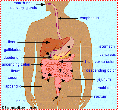 The human digestive system is a complex series of organs and glands that processes food. In order to use the food we eat, our body has to break the food down into smaller molecules that it can process; it also has to excrete waste.
 Most of the digestive organs (like the stomach and intestines) are tube-like and contain the food as it makes its way through the body. The digestive system is essentially a long, twisting tube that runs from the mouth to the anus, plus a few other organs (like the liver and pancreas) that produce or store digestive chemicals.
The Digestive Process:
The start of the process - the mouth: The digestive process begins in the mouth. Food is partly broken down by the process of chewing and by the chemical action of salivary enzymes (these enzymes are produced by the salivary glands and break down starches into smaller molecules).
On the way to the stomach: the esophagus - After being chewed and swallowed, the food enters the esophagus. The esophagus is a long tube that runs from the mouth to the stomach. It uses rhythmic, wave-like muscle movements (called peristalsis) to force food from the throat into the stomach. This muscle movement gives us the ability to eat or drink even when we're upside-down.
In the stomach - The stomach is a large, sack-like organ that churns the food and bathes it in a very strong acid (gastric acid). Food in the stomach that is partly digested and mixed with stomach acids is called chyme.
In the small intestine - After being in the stomach, food enters the duodenum, the first part of the small intestine. It then enters the jejunum and then the ileum (the final part of the small intestine). In the small intestine, bile (produced in the liver and stored in the gall bladder), pancreatic enzymes, and other digestive enzymes produced by the inner wall of the small intestine help in the breakdown of food.
In the large intestine - After passing through the small intestine, food passes into the large intestine. In the large intestine, some of the water and electrolytes (chemicals like sodium) are removed from the food. Many microbes (bacteria like Bacteroides, Lactobacillus acidophilus, Escherichia coli, and Klebsiella) in the large intestine help in the digestion process. The first part of the large intestine is called the cecum (the appendix is connected to the cecum). Food then travels upward in the ascending colon. The food travels across the abdomen in the transverse colon, goes back down the other side of the body in the descending colon, and then through the sigmoid colon.
The end of the process - Solid waste is then stored in the rectum until it is excreted via the anus.
Digestive System Glossary:
anus - the opening at the end of the digestive system from which feces (waste) exits the body.
appendix - a small sac located on the cecum.
ascending colon - the part of the large intestine that run upwards; it is located after the cecum.
bile - a digestive chemical that is produced in the liver, stored in the gall bladder, and secreted into the small intestine.
cecum - the first part of the large intestine; the appendix is connected to the cecum.
chyme - food in the stomach that is partly digested and mixed with stomach acids. Chyme goes on to the small intestine for further digestion.
descending colon - the part of the large intestine that run downwards after the transverse colon and before the sigmoid colon.
duodenum - the first part of the small intestine; it is C-shaped and runs from the stomach to the jejunum.
epiglottis - the flap at the back of the tongue that keeps chewed food from going down the windpipe to the lungs. When you swallow, the epiglottis automatically closes. When you breathe, the epiglottis opens so that air can go in and out of the windpipe.
esophagus - the long tube between the mouth and the stomach. It uses rhythmic muscle movements (called peristalsis) to force food from the throat into the stomach.
gall bladder - a small, sac-like organ located by the duodenum. It stores and releases bile (a digestive chemical which is produced in the liver) into the small intestine.
ileum - the last part of the small intestine before the large intestine begins.
jejunum - the long, coiled mid-section of the small intestine; it is between the duodenum and the ileum.
liver - a large organ located above and in front of the stomach. It filters toxins from the blood, and makes bile (which breaks down fats) and some blood proteins.
mouth - the first part of the digestive system, where food enters the body. Chewing and salivary enzymes in the mouth are the beginning of the digestive process (breaking down the food).
pancreas - an enzyme-producing gland located below the stomach and above the intestines. Enzymes from the pancreas help in the digestion of carbohydrates, fats and proteins in the small intestine.
peristalsis - rhythmic muscle movements that force food in the esophagus from the throat into the stomach. Peristalsis is involuntary - you cannot control it. It is also what allows you to eat and drink while upside-down.
rectum - the lower part of the large intestine, where feces are stored before they are excreted.
salivary glands - glands located in the mouth that produce saliva. Saliva contains enzymes that break down carbohydrates (starch) into smaller molecules.
sigmoid colon - the part of the large intestine between the descending colon and the rectum.
stomach - a sack-like, muscular organ that is attached to the esophagus. Both chemical and mechanical digestion takes place in the stomach. When food enters the stomach, it is churned in a bath of acids and enzymes.
transverse colon - the part of the large intestine that runs horizontally across the abdomen.
Goals
- Restoration of fluid and electrolyte balance.
- Client will express feelings of self-worth.
- Adequate nutritional status will be maintained.
- Client will experience decreased frequency of regular bowel habits.
- Client will establish regular bowel habits of appropriate amount and consistency.
- Client will be free from pain.
- Effective breathing patterns will be maintained.
- Effective communication methods will be established.
- Skin integrity will be restored/maintained.
Interventions
Enemas
- General information
- Instillation of fluid into the rectum, usually for the purpose of stimulating defecation. The various types include
- cleansing enema (tap water, normal saline, or soap): used to treat constipation or feces impaction, as bowel cleansing prior to diagnostic procedures or surgery, to help establish regular bowel functions.
- retention enema (mineral oil, olive oil, cottonseed oil): usually administered to lubricate or soften a hard fecal mass to facilitate defecation.
- Nursing care for a cleansing enema
- Explain procedure and that breathing through the mouth relaxes abdominal musculature and helps to avoid cramps; explain the need to take adequate time to defecate.
- Assemble equipment: prepare solution at 105°-110°F and have bedpan, commode, or nearby bathroom ready for use.
- Position client and drape adequately.
- Place waterproof pad under buttocks.
- Lubricate tube and allow solution to fill the tubing, displacing air.
- Insert rectal tube 4-5 inches without using force; request that client take several deep breaths.
- Administer 500-100 ml of solution over 5-10 minutes; if cramping occurs slow the speed of instillation.
- After administration, have the client retain solution until the urge to defecate becomes strong.
- Document amount, color, characteristics of stool, and client's reaction during procedure.
- Assess for dizziness, light-headedness, abdominal cramps, nausea.
- Monitor electrolyte levels if client is to receive repeated enemas.
- Nursing care for a retention enema: same as for a cleansing enema except
- Oil is used instead of water (comes prepared in commercial kits and is given at body temperature).
- Administer 150-200 ml of prepared solution.
- Instruct client to retain oil for at least 30 minutes in order for it to take effect.
Gastrostomy
- General information
- Insertion of a catheter through an abdominal incision into the stomach where it is secured with sutures.
- Used as an alternative method of feeding, either temporary or permanent, for clients who have problems with swallowing, ingestion, and digestion.
- Nursing care
- Maintain skin integrity: inspect and cleanse skin around stoma frequently; keep deep area dry to avoid excoriation.
- Maintain patency of the gastrostomy tube.
- Assess for residual before each feeding (check orders concerning withholding feeding).
- Irrigate tube before and after meals.
- Measure/record any drainage.
- Promote adequate nutrition.
- Administer feeding with client in high-Fowler's and keep head of bed elevated for 30 minutes after meals to prevent regurgitation.
- Maintain feeding at room temperature.
- Ensure that prescribed amount of feeding be given within prescribed amount of time.
- Weigh client daily.
- Monitor I&O until feedings are well tolerated.
- Monitor for signs of dehydration.
Nasogastric (NG) Tubes
- General information
- Soft rubber or plastic tube inserted through a nostril and into the stomach for gastric decompression, feeding, or obtaining specimens for analysis of stomach contents
- Types
- Levin: single-lumen, nonvented
- Nursing care
- Insertion of the tube
- Explain the purpose of the tube and the procedure for insertion.
- Measure the tube: distance on the tube from the tip of the nose to the ear lobe plus the distance from the ear lobe to the tip of the xiphoid.
- Instruct client to bend head forward if possible during insertion.
- Monitor functioning of system and ensure patency of the NG tube: abdominal discomfort/distension, nausea and vomiting, and little or no drainage in collection bottle are all signs that system is not functioning properly.
- Assess the position: aspirate gastric contents to confirm that tube is in stomach; inject 10 ml air through tube and auscultate for rapid influx.
- Check that tubing is free of kinks; irrigate as per physician order.
- Record amount, color, and odor of drainage.
- Provide measures to ensure maximal comfort.
- Apply water-soluble lubricant to lips to prevent dryness.
- Keep nares free from secretions.
- Provide periodic warm saline gargles to prevent dryness.
- Provide frequent mouth care with toothbrush/toothpaste or flavored mouthwashes.
- If allowed, give client hard candy or gum to stimulate the flow of saliva and prevent dryness.
- Elevate head and chest during and for 1-2 hours after feedings to prevent reflux (most comfortable position when suction is used).
- Monitor/maintain fluid and electrolyte balance.
- Assess for signs of metabolic alkalosis (suctioning causes excessive loss of hydrochloric acid and potassium).
- Administer IV fluids as ordered.
- If suction used, irrigate NG tube with normal saline to decrease sodium loss.
- Keep accurate I&O.
- If suction used provide ice chips sparingly (if allowed) to avoid dilution of electrolytes.
- Monitor lab values and electrolytes frequently.
Intestinal Tubes
- General information
- Tube is inserted via a nostril through the stomach and into the intestine for decompression proximal to an obstruction, relief of an obstruction, decompression of post-op edema at the surgical site.
- Types
- Cantor tube: single lumen
- Harris tube: single lumen
- Miller-Abbott: double lumen
- Nursing care
- Facilitate placement of the tube.
- Position client in high-Fowler's while tube is being passed from the nose to the stomach; then place client on right side to aid in advancing the tube from the stomach to duodenum.
- Continuously monitor tube markings.
- Tape tube in place only after placement in duodenum is confirmed.
- Provide measures for maximal comfort, as for NG tube.


