

DIGESTIVE SYSTEM
| Human Anatomy | |
 The human digestive system is a complex series of organs and glands that processes food. In order to use the food we eat, our body has to break the food down into smaller molecules that it can process; it also has to excrete waste.
 Most of the digestive organs (like the stomach and intestines) are tube-like and contain the food as it makes its way through the body. The digestive system is essentially a long, twisting tube that runs from the mouth to the anus, plus a few other organs (like the liver and pancreas) that produce or store digestive chemicals.
The Digestive Process:
The start of the process - the mouth: The digestive process begins in the mouth. Food is partly broken down by the process of chewing and by the chemical action of salivary enzymes (these enzymes are produced by the salivary glands and break down starches into smaller molecules).
On the way to the stomach: the esophagus - After being chewed and swallowed, the food enters the esophagus. The esophagus is a long tube that runs from the mouth to the stomach. It uses rhythmic, wave-like muscle movements (called peristalsis) to force food from the throat into the stomach. This muscle movement gives us the ability to eat or drink even when we're upside-down.
In the stomach - The stomach is a large, sack-like organ that churns the food and bathes it in a very strong acid (gastric acid). Food in the stomach that is partly digested and mixed with stomach acids is called chyme.
In the small intestine - After being in the stomach, food enters the duodenum, the first part of the small intestine. It then enters the jejunum and then the ileum (the final part of the small intestine). In the small intestine, bile (produced in the liver and stored in the gall bladder), pancreatic enzymes, and other digestive enzymes produced by the inner wall of the small intestine help in the breakdown of food.
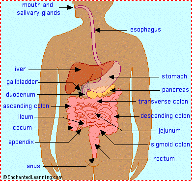
In the large intestine - After passing through the small intestine, food passes into the large intestine. In the large intestine, some of the water and electrolytes (chemicals like sodium) are removed from the food. Many microbes (bacteria like Bacteroides, Lactobacillus acidophilus, Escherichia coli, and Klebsiella) in the large intestine help in the digestion process. The first part of the large intestine is called the cecum (the appendix is connected to the cecum). Food then travels upward in the ascending colon. The food travels across the abdomen in the transverse colon, goes back down the other side of the body in the descending colon, and then through the sigmoid colon.
The end of the process - Solid waste is then stored in the rectum until it is excreted via the anus.
Digestive System Glossary:
anus - the opening at the end of the digestive system from which feces (waste) exits the body.
appendix - a small sac located on the cecum.
ascending colon - the part of the large intestine that run upwards; it is located after the cecum.
bile - a digestive chemical that is produced in the liver, stored in the gall bladder, and secreted into the small intestine.
cecum - the first part of the large intestine; the appendix is connected to the cecum.
chyme - food in the stomach that is partly digested and mixed with stomach acids. Chyme goes on to the small intestine for further digestion.
descending colon - the part of the large intestine that run downwards after the transverse colon and before the sigmoid colon.
duodenum - the first part of the small intestine; it is C-shaped and runs from the stomach to the jejunum.
epiglottis - the flap at the back of the tongue that keeps chewed food from going down the windpipe to the lungs. When you swallow, the epiglottis automatically closes. When you breathe, the epiglottis opens so that air can go in and out of the windpipe.
esophagus - the long tube between the mouth and the stomach. It uses rhythmic muscle movements (called peristalsis) to force food from the throat into the stomach.
gall bladder - a small, sac-like organ located by the duodenum. It stores and releases bile (a digestive chemical which is produced in the liver) into the small intestine.
ileum - the last part of the small intestine before the large intestine begins.
jejunum - the long, coiled mid-section of the small intestine; it is between the duodenum and the ileum.
liver - a large organ located above and in front of the stomach. It filters toxins from the blood, and makes bile (which breaks down fats) and some blood proteins.
mouth - the first part of the digestive system, where food enters the body. Chewing and salivary enzymes in the mouth are the beginning of the digestive process (breaking down the food).
pancreas - an enzyme-producing gland located below the stomach and above the intestines. Enzymes from the pancreas help in the digestion of carbohydrates, fats and proteins in the small intestine.
peristalsis - rhythmic muscle movements that force food in the esophagus from the throat into the stomach. Peristalsis is involuntary - you cannot control it. It is also what allows you to eat and drink while upside-down.
rectum - the lower part of the large intestine, where feces are stored before they are excreted.
salivary glands - glands located in the mouth that produce saliva. Saliva contains enzymes that break down carbohydrates (starch) into smaller molecules.
sigmoid colon - the part of the large intestine between the descending colon and the rectum.
stomach - a sack-like, muscular organ that is attached to the esophagus. Both chemical and mechanical digestion takes place in the stomach. When food enters the stomach, it is churned in a bath of acids and enzymes.
transverse colon - the part of the large intestine that runs horizontally across the abdomen.
Goals
- Restoration of fluid and electrolyte balance.
- Client will express feelings of self-worth.
- Adequate nutritional status will be maintained.
- Client will experience decreased frequency of regular bowel habits.
- Client will establish regular bowel habits of appropriate amount and consistency.
- Client will be free from pain.
- Effective breathing patterns will be maintained.
- Effective communication methods will be established.
- Skin integrity will be restored/maintained.
Interventions
Enemas
- General information
- Instillation of fluid into the rectum, usually for the purpose of stimulating defecation. The various types include
- cleansing enema (tap water, normal saline, or soap): used to treat constipation or feces impaction, as bowel cleansing prior to diagnostic procedures or surgery, to help establish regular bowel functions.
- retention enema (mineral oil, olive oil, cottonseed oil): usually administered to lubricate or soften a hard fecal mass to facilitate defecation.
- Nursing care for a cleansing enema
- Explain procedure and that breathing through the mouth relaxes abdominal musculature and helps to avoid cramps; explain the need to take adequate time to defecate.
- Assemble equipment: prepare solution at 105°-110°F and have bedpan, commode, or nearby bathroom ready for use.
- Position client and drape adequately.
- Place waterproof pad under buttocks.
- Lubricate tube and allow solution to fill the tubing, displacing air.
- Insert rectal tube 4-5 inches without using force; request that client take several deep breaths.
- Administer 500-100 ml of solution over 5-10 minutes; if cramping occurs slow the speed of instillation.
- After administration, have the client retain solution until the urge to defecate becomes strong.
- Document amount, color, characteristics of stool, and client's reaction during procedure.
- Assess for dizziness, light-headedness, abdominal cramps, nausea.
- Monitor electrolyte levels if client is to receive repeated enemas.
- Nursing care for a retention enema: same as for a cleansing enema except
- Oil is used instead of water (comes prepared in commercial kits and is given at body temperature).
- Administer 150-200 ml of prepared solution.
- Instruct client to retain oil for at least 30 minutes in order for it to take effect.
Gastrostomy
- General information
- Insertion of a catheter through an abdominal incision into the stomach where it is secured with sutures.
- Used as an alternative method of feeding, either temporary or permanent, for clients who have problems with swallowing, ingestion, and digestion.
- Nursing care
- Maintain skin integrity: inspect and cleanse skin around stoma frequently; keep deep area dry to avoid excoriation.
- Maintain patency of the gastrostomy tube.
- Assess for residual before each feeding (check orders concerning withholding feeding).
- Irrigate tube before and after meals.
- Measure/record any drainage.
- Promote adequate nutrition.
- Administer feeding with client in high-Fowler's and keep head of bed elevated for 30 minutes after meals to prevent regurgitation.
- Maintain feeding at room temperature.
- Ensure that prescribed amount of feeding be given within prescribed amount of time.
- Weigh client daily.
- Monitor I&O until feedings are well tolerated.
- Monitor for signs of dehydration.
Nasogastric (NG) Tubes
- General information
- Soft rubber or plastic tube inserted through a nostril and into the stomach for gastric decompression, feeding, or obtaining specimens for analysis of stomach contents
- Types
- Levin: single-lumen, nonvented
- Nursing care
- Insertion of the tube
- Explain the purpose of the tube and the procedure for insertion.
- Measure the tube: distance on the tube from the tip of the nose to the ear lobe plus the distance from the ear lobe to the tip of the xiphoid.
- Instruct client to bend head forward if possible during insertion.
- Monitor functioning of system and ensure patency of the NG tube: abdominal discomfort/distension, nausea and vomiting, and little or no drainage in collection bottle are all signs that system is not functioning properly.
- Assess the position: aspirate gastric contents to confirm that tube is in stomach; inject 10 ml air through tube and auscultate for rapid influx.
- Check that tubing is free of kinks; irrigate as per physician order.
- Record amount, color, and odor of drainage.
- Provide measures to ensure maximal comfort.
- Apply water-soluble lubricant to lips to prevent dryness.
- Keep nares free from secretions.
- Provide periodic warm saline gargles to prevent dryness.
- Provide frequent mouth care with toothbrush/toothpaste or flavored mouthwashes.
- If allowed, give client hard candy or gum to stimulate the flow of saliva and prevent dryness.
- Elevate head and chest during and for 1-2 hours after feedings to prevent reflux (most comfortable position when suction is used).
- Monitor/maintain fluid and electrolyte balance.
- Assess for signs of metabolic alkalosis (suctioning causes excessive loss of hydrochloric acid and potassium).
- Administer IV fluids as ordered.
- If suction used, irrigate NG tube with normal saline to decrease sodium loss.
- Keep accurate I&O.
- If suction used provide ice chips sparingly (if allowed) to avoid dilution of electrolytes.
- Monitor lab values and electrolytes frequently.
Intestinal Tubes
- General information
- Tube is inserted via a nostril through the stomach and into the intestine for decompression proximal to an obstruction, relief of an obstruction, decompression of post-op edema at the surgical site.
- Types
- Cantor tube: single lumen
- Harris tube: single lumen
- Miller-Abbott: double lumen
- Nursing care
- Facilitate placement of the tube.
- Position client in high-Fowler's while tube is being passed from the nose to the stomach; then place client on right side to aid in advancing the tube from the stomach to duodenum.
- Continuously monitor tube markings.
- Tape tube in place only after placement in duodenum is confirmed.
- Provide measures for maximal comfort, as for NG tube.


0 comments:
Post a Comment